 ultimate-guide
Effective Treatments for Herniated Discs: 2026 Guide
Table of Contents
- What a Herniated Disc Is and Why It Hurts
- Symptoms That Point to a Herniated Disc
- Effective Treatments for Herniated Discs Without Surgery
- Non-Surgical Spinal Decompression: How It Works
- Herniated Disc Recovery Timeline: What to Expect Week by Week
- When Is Surgery Necessary for a Herniated Disc?
- Ergonomics, Nutrition, and Mental Health: The Recovery Factors Most People Skip
- Frequently Asked Questions
Last Updated: September 19, 2026
What a Herniated Disc Is and Why It Hurts
A herniated disc is a spinal injury in which the soft inner core of a vertebral disc pushes through a tear in its tougher outer ring, pressing on nearby nerve roots. That pressure, plus the inflammation it triggers, produces the shooting pain, numbness, and weakness that define the condition.
Bulging Disc vs. Herniated Disc: What the MRI Shows
A bulging disc extends evenly beyond its normal boundary without a tear in the outer ring. A herniated disc involves an actual rupture, with inner material escaping through the annulus. On an MRI, a bulge looks like a smooth, broad protrusion; a herniation appears as a focal outpouching, often at the lumbar or cervical level.
Symptoms That Point to a Herniated Disc
The signature symptom is pain that travels along a nerve pathway rather than staying in one spot. In the lower back, that means sciatica: pain radiating from the buttock down the leg, sometimes past the knee. In the neck, it means pain shooting into the shoulder, arm, or hand.
Watch for these additional signs:
- Numbness or tingling in the arms, hands, legs, or feet
- Muscle weakness that makes lifting or gripping harder than usual
- Pain that worsens with sitting, coughing, sneezing, or bending forward
- A deep ache near the spine that improves when you lie down
- Reduced reflexes on one side of the body
Effective Treatments for Herniated Discs Without Surgery
Most herniated discs respond to conservative treatment. The American Academy of Orthopaedic Surgeons notes that the majority of patients improve within six to twelve weeks without an operation, which is why nonsurgical care is almost always the first step.
Medication, Injections, and Pain Management
Pain management buys the time your disc needs to heal. NSAIDs reduce pain and the inflammation irritating the nerve, muscle relaxants ease spasm, and when oral medication is not enough, corticosteroid injections deliver anti-inflammatory medication directly to the inflamed nerve root.
| Approach | What It Does | Typical Role |
|---|---|---|
| NSAIDs | Reduce pain and inflammation | First-line, short-term |
| Muscle relaxants | Calm muscle spasm | Short-term relief |
| Corticosteroid injections | Target nerve-root inflammation | When oral meds fall short |
| Physical therapy | Restore strength and mobility | Core of recovery |
Physical therapy, Heat, and Cold
Physical therapy is the backbone of recovery. A therapist builds a program around core stabilization, posture correction, and gentle stretching that takes pressure off the affected nerve root. Heat relaxes tight muscles before activity; cold reduces inflammation after. Neither replaces exercise, but both make movement more comfortable early on.
Non-Surgical Spinal Decompression: How It Works
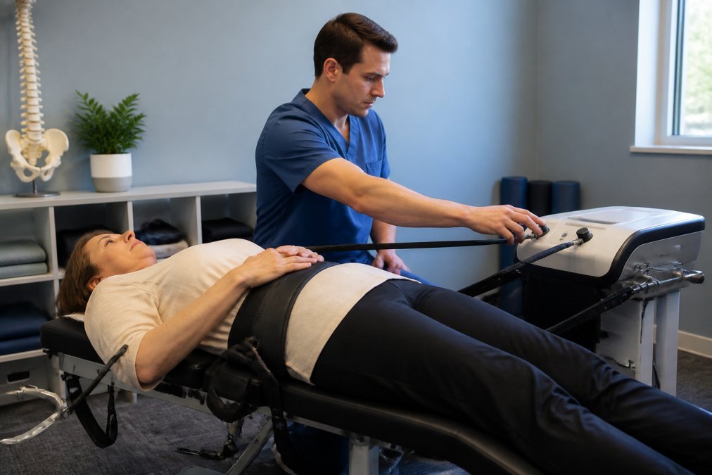
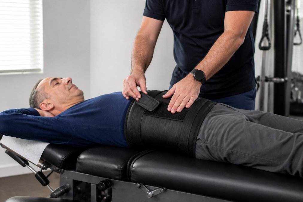
Non-surgical spinal decompression uses a motorized table to apply controlled, gently varying traction to the spine, creating negative pressure inside the disc that may help a herniation retract and draw nutrients back into the tissue. Sessions typically last 30 to 45 minutes and repeat over several weeks.
Herniated Disc Recovery Timeline: What to Expect Week by Week
Recovery from a herniated disc is less a straight line than a series of phases, each with its own goals and traps. The arc below reflects the pattern most clinicians see, but the size and location of the herniation, your age, fitness, and how quickly you start appropriate care all shift the pace.
Weeks 1-2: Calm the Nerve
This is the peak-pain phase: reduce inflammation, protect the nerve root, and avoid positions that provoke symptoms.
- Activity: Relative rest, not bed rest, prolonged lying down weakens supporting muscles. Short, frequent walks on flat ground are usually better tolerated than sitting.
- Positioning: Lumbar herniations often feel worse with prolonged sitting and bending forward; cervical herniations with neck extension and overhead reaching. Find positions that reduce radiating pain and use them deliberately.
- Modalities: Ice for the first 48-72 hours after an acute flare, then heat for muscle spasm. Neither treats the disc itself, but both make movement tolerable.
- Medication: NSAIDs and, if prescribed, muscle relaxants. Follow dosing instructions and check with a clinician if you have kidney, stomach, or cardiovascular conditions.
Weeks 3-6: Restore Gentle Motion
Pain typically begins to ease, but nerve symptoms can still flare with specific movements. This is when physical therapy usually starts in earnest.
- Therapy focus: Core stabilization, directional preference exercises (McKenzie-style), posture correction, and graded walking. A therapist identifies which movements centralize your pain, moving it from the leg or arm back toward the spine, and builds the program around those.
- What to avoid: Heavy lifting, twisting under load, and any activity that reproduces shooting pain down the limb. Local back or neck discomfort is generally more tolerable than radiating pain.
- Realistic expectation: Many people notice meaningful improvement by week four to six. If you do not, that is information, not failure.
Weeks 6-12: The Decision Fork
Six weeks is the conventional checkpoint. Most herniated discs that will respond to conservative care have shown clear signs by now.
Call for a FREE MRI Review and Consultation! →
- If you are improving: Expand strength and mobility work. Add resistance training for the core, hips, and upper back under a therapist's guidance. Aerobic activity, walking, stationary cycling, swimming, supports healing and mood.
- If you are plateaued: Reassess with imaging (MRI if not already done), a specialist evaluation, and a candid conversation about escalation. Options include epidural steroid injections, a trial of non-surgical spinal decompression, or surgical consultation in selected cases.
- If you are worsening: Do not wait out the next six weeks. Worsening neurological findings change the calculus.
Months 3-6: Consolidate and Prevent Recurrence
For most people, residual numbness or tingling fades during this window, and full function returns. The work shifts from symptom relief to durability.
- Strength: Progressive loading of the posterior chain, core, and scapular stabilizers.
- Habits: Ergonomic setup, lifting mechanics, and consistent movement breaks.
- Warning signs: A recurrence of radiating pain, new weakness, or numbness returning after improvement warrants re-evaluation rather than a wait-and-see approach.
Red Flags That Override the Timeline
Certain symptoms mean the schedule above no longer applies. Seek urgent evaluation for:
- Loss of bladder or bowel control
- Numbness in the saddle region (inner thighs, groin, perineum)
- Rapidly progressive weakness in an arm or leg
- Severe pain that is unrelenting regardless of position
When Is Surgery Necessary for a Herniated Disc?
Surgery is the right answer for a minority of herniated discs. The question is not whether surgery works, it often does, but whether the situation justifies the risks, cost, and recovery time compared with continuing conservative care.
The Three Clear Indications
Most spine surgeons agree on a narrow set of situations where surgery moves from optional to advisable:
- Progressive neurological deficit. Measurable weakness that is getting worse over days to weeks, not better. This is the most time-sensitive indication because nerve damage can become permanent.
- Cauda equina syndrome. Loss of bladder or bowel control, saddle numbness, or rapidly worsening leg weakness. This is a surgical emergency and requires immediate evaluation.
- Severe, persistent pain after a thorough conservative course. Typically defined as six to twelve weeks of structured non-surgical care, physical therapy, medication, and often injections, without meaningful improvement, and with pain that is significantly limiting function.
The Procedures, and What Each One Actually Does
- Microdiscectomy / discectomy. The most common operation: the surgeon removes the portion of the disc pressing on the nerve root. Microdiscectomy uses a smaller incision and a microscope or endoscope. It is typically outpatient, and many patients get significant leg or arm pain relief within days, though back or neck soreness lingers longer.
- Laminectomy. Removal of part of the vertebral bone (the lamina) to widen the spinal canal. More often used for spinal stenosis than a simple herniation, but it may be combined with discectomy when the canal is narrow.
- Spinal fusion. Permanently joins two vertebrae using bone graft, hardware, or both. Reserved for instability, recurrent herniation, or cases where removing disc material would destabilize the spine. Fusion has a longer recovery and changes adjacent-segment mechanics, so it is not first-line for a straightforward herniation.
- Artificial disc replacement. An alternative to fusion in carefully selected patients, typically cervical or lumbar. It preserves motion at the operated level, which may reduce stress on adjacent segments. Not every patient is a candidate, and long-term outcomes depend on proper selection.
- Minimally invasive approaches. Smaller incisions, less muscle disruption, and often shorter hospital stays. They are not automatically better for every case, the right approach depends on the anatomy and the surgeon's experience.
What Post-Surgery Rehabilitation Actually Looks Like
This is where most guides get vague, and where patients most often feel unprepared.
- First 24-72 hours: Usually walking within hours of surgery. Instructions typically limit bending, twisting, and lifting (often nothing heavier than a gallon of milk) for the first couple of weeks.
- Weeks 1-2: Short, frequent walks, incision care, and pain medication as needed. Many people return to desk work within one to two weeks, depending on the procedure and their commute.
- Weeks 2-6: Formal physical therapy usually begins. Early goals are gentle range of motion, walking endurance, and core activation, not heavy strengthening. Lifting restrictions typically remain.
- Weeks 6-12: Progressive strengthening and a gradual return to more demanding activity. Some patients are cleared for light sport or recreation in this window; others are not.
- Months 3-6: Most patients return to normal activities. Full recovery, including heavy lifting or high-impact sport, often takes longer and depends on the procedure.
Risks and Trade-Offs Worth Knowing
No operation is risk-free. For discectomy, the most discussed risks are reherniation (which can require a second operation), infection, nerve injury, and incomplete pain relief. Fusion carries additional risks related to hardware and adjacent-segment degeneration over time. These risks are generally low in experienced hands, but they are real, and they are the reason surgery is reserved for cases where the conservative path has failed or a red flag is present.
Ergonomics, Nutrition, and Mental Health: The Recovery Factors Most People Skip
Three factors shape recovery as much as any treatment, and most guides ignore them.
Frequently Asked Questions
Can a herniated disc heal without surgery?
Many herniated discs improve with conservative care. A common approach combines rest, anti-inflammatory medication, physical therapy, and non-surgical spinal decompression. Symptoms often ease over several weeks as inflammation around the nerve root settles, even though the disc itself may not fully return to its original shape. If pain, numbness, or muscle weakness gets worse, or you lose bladder or bowel control, seek medical care right away.
How long does the herniated disc recovery timeline usually take?
Most people notice meaningful improvement within four to six weeks of starting conservative treatment, with continued gains over three to six months. The herniated disc recovery timeline depends on the location (lumbar vs. cervical), the size of the herniation, your age, and how consistently you follow your treatment plan. A smaller bulge with mild tingling typically resolves faster than a large extrusion causing radiculopathy and muscle weakness.
When is surgery necessary for a herniated disc?
Surgery is usually considered when conservative treatment has not helped after six to twelve weeks, when imaging shows a large herniation pressing on a nerve root, or when there is a neurological deficit such as progressive muscle weakness or loss of bladder or bowel control. Procedures range from minimally invasive microdiscectomy to laminectomy or spinal fusion. Your physician can review your MRI and symptoms to decide if surgery is appropriate.
Is non-surgical spinal decompression different from regular chiropractic adjustment?
Yes. A chiropractic adjustment applies a quick, targeted thrust to restore joint motion. Non-surgical spinal decompression uses a motorized table to apply a gentle, controlled stretch to the spine over several minutes, which can create negative pressure inside the disc. That negative pressure may help a bulging or herniated disc retract and allow nutrients to flow back into the disc. Many patients combine both approaches during recovery.